Invented by Japanese specialists in ultrasound, sonoelastography is a new technique that measures the quality and quantity of mammary tumors elasticity. Using sonoelastography removes many of the difficulties of breast cancer diagnosis, because it combines the advantages of ultrasound (depth of penetration and resolution) with sensitivity to differences in stiffness of the analyzed tissue.
The technique is based on the fact that malignant tissue is harder than benign tissue, and measure the elastic properties using ultrasound. This method can detect and locate tumors when they have a different elasticity than the surrounding tissue, complementing mammography, ultrasound two-dimensional nuclear magnetic resonance and Doppler examination.

Elasticity of cancerous tumors is reduced from the one of the noncancerous tumors, so the method can distinguish, with accuracy that reaches 90%, malignant tumors (cancerous) from benign (noncancerous). This type of ultrasound tecnhinque is applicable to small tumors, less than 2 cm, which implies an early diagnosis of breast cancer, which, with early treatment the rate of healing the disease is much higher.
For tumors of any size, sonoelastography has a specificity of 93% and a sensitivity of 80%, positive predictive power of 85.3% and negative predictive power of 90.3%. The sensitivity is better for lesions less than 5 mm, and specificity for lesions over 10 mm in size.
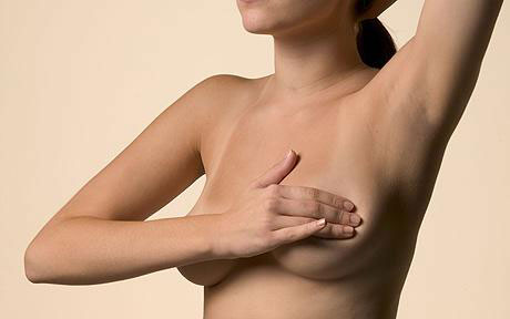
Sonoelastography uses low-frequency transverse amplitude which is propagated through internal organs to measure the elastic properties of a tissue. For investigation, doctors use a special probe to scan the breast. The examination result is interpreted as a color scale from red to blue, numbered from one to five, depending on the severity of the disease.
Red indicates a high elasticity of the breast. A low elasticity means the cancer tumor and the predominant color is deep blue. One of the advantages of this investigation is the lack of pain. In addition, it is non-invasive from breast, without irradiation or other side effects. Furthermore, the method can be used for other medical tests at the level of soft tissue and difficult to assess: thyroid, prostate, pancreas, liver, vascular and musculoskeletal system level.